Chiropractic and Health Books
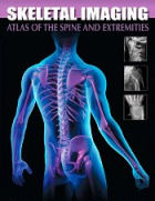
Skeletal Imaging: Atlas of the Spine and Extremities
 Skeletal Imaging: Atlas of the Spine and Extremities
Check New and Used Prices
Use this atlas to accurately interpret images of musculoskeletal disorders! Taylor, Hughes, and Resnick's Skeletal Imaging: Atlas of the Spine and Extremities, 2nd Edition covers each anatomic region separately, so common disorders are shown within the context of each region. This allows you to examine and compare images for a variety of different disorders. A separate chapter is devoted to each body region, with coverage of normal developmental anatomy, developmental anomalies and normal variations, and how to avoid a misdiagnosis by differentiating between disorders that appear to be similar. All of the most frequently encountered musculoskeletal conditions are included, from physical injuries to tumors to infectious diseases.
Key Features
*Over 2,100 images include radiographs, radionuclide studies, CT scans, and MR images, illustrating pathologies and comparing them with other disorders in the same region.
*Organization by anatomic region addresses common afflictions for each region in separate chapters, so you can see how a particular region looks when affected by one condition as compared to its appearance with other conditions.
*Coverage of each body region includes normal developmental anatomy, fractures, deformities, dislocations, infections, hematologic disorders, and more.
*Normal Developmental Anatomy sections open each chapter, describing important developmental landmarks in various regions of the body from birth to skeletal maturity.
*Practical tables provide a quick reference to essential information, including normal developmental anatomic milestones, developmental anomalies, common presentations and symptoms of diseases, and much more.
Table of Contents
Part I Introduction
1. Introduction to Skeletal Disorders: General Concepts
Part II Spine
2. Cervical Spine
3. Thoracic Spine
4. Lumbar Spine
5. Sacrococcygeal Spine and Sacroiliac Joints
Part III Pelvis and Lower Extremities
6. Pelvis and Symphysis Pubis
7. Hip
8. Femur
9. Knee
10. Tibia and Fibula
11. Ankle and Foot
Part IV Thoracic Cage and Upper Extremities
12. Ribs, Sternum, and Sternoclavicular Joints
13. Clavicle, Scapula, and Shoulder
14. Humerus
15. Elbow
16. Radius and Ulna
17. Wrist and Hand
Skeletal Imaging: Atlas of the Spine and Extremities
Check New and Used Prices